Diagnosing and Staging Lung Cancer
If your healthcare provider thinks you may have lung cancer, a number of tests might be done to be sure. Testing is needed to diagnose lung cancer and find out the type of lung cancer, where it is, and if, or how much, it has spread. Test results also help your healthcare provider plan treatment.
What do the tests show?
Each test result for lung cancer offers a piece of information. Taken as a whole, the results can give many details about your cancer. For instance, do you have non-small cell or small cell lung cancer? What type of non-small lung cell cancer or type of small cell lung cancer? How big is the tumor? Where is the tumor? Are there lymph nodes involved? Has the cancer spread? If so, where? With these details, you and your healthcare provider can start to plan treatment.
Biopsy
In a biopsy, a small piece (called a sample) of tissue is removed. It can be taken from a tumor in the lung. Or it can be taken from tumors in other parts of your body if the cancer has spread outside of the lung and a lung biopsy can't be done. The sample is sent to a lab where it's tested to learn more about your cancer. A biopsy is the only way to know for sure if you have lung cancer. These tests can be used to get a biopsy:
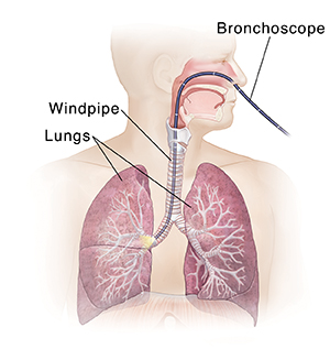 |
| A bronchoscope provides a direct view of the windpipe and bronchial tubes. |
-
Bronchoscopy. A thin, lighted, flexible tube (called a bronchoscope) is put into your nose and down your windpipe. The healthcare provider then uses the scope to look at your windpipe and main breathing (bronchial) tubes. Tissue samples may be taken from any areas that look like they might be cancer. This can be done with a tool that's used within the bronchoscope.
-
Mediastinoscopy. A thin, lighted tube is put through a small cut (incision) above the sternum (breastbone). It's used to look at the space between the lungs. A biopsy can be done through this tube. A mediastinotomy works much the same, but the tube is put in between the ribs through an incision in the chest wall. This allows your healthcare provider to look at and biopsy lymph nodes and other tissues in your chest.
-
Endobronchial ultrasound. A bronchoscope is used with ultrasound (images made from sound waves) to look at the lymph nodes and other structures between the lungs. Another test like this is called an endoscopic esophageal ultrasound. In that test, a scope is passed down the tube that carries food from your mouth to your stomach (the esophagus) to look at nearby lymph nodes. If any of the lymph nodes look swollen, a sample of tissue cells will be taken out and tested for cancer cells. This is done with a needle that's used through the scope.
-
Core-needle biopsy. A thin needle is used to remove a core of tissue from a tumor. This test is done under local anesthesia to help prevent pain. This means medicines are used to numb the area the needle passes through. During the core needle biopsy, a CT scan may be used to help the healthcare provider put the needle exactly where it needs to be.
Imaging tests
Pictures from different imaging tests can give details about your lungs and any tumors in them. These pictures can also show exactly where a tumor is and how big it is.
-
Chest X-ray. This is one of the most common imaging tests. It can show changes in and around your lungs.
-
CT scan. This scan takes many X-ray images from different angles. A computer puts them together to make detailed 3-D images. The CT scan will be done of your chest, abdomen, and pelvis. You may receive a contrast medium by mouth or injected into your vein before the test. The contrast helps make the pictures clearer.
-
PET scan (positron emission tomography). This test uses a slightly radioactive liquid sugar (tracer). It's put into your blood and absorbed by cancer cells anywhere in your body. The scanner then checks your whole body for places where the tracer has collected. A PET scan is often done along with a CT scan. This is called a PET/CT scan and can find small cancers that can't be found by CT scan alone.
-
MRI. This test is not used often to look at the lungs. It may be used to check for cancer spread to your brain or bones. MRI uses strong magnets and computers to form a highly detailed image.
You may also have a bone scan or other imaging tests to learn whether the cancer has spread. Your healthcare provider will tell you how to prepare for any tests and what the test will be like.
Staging lung cancer
Staging is a process to measure how much cancer there is and whether it has spread. Your healthcare provider uses exams and tests to find out the size of the cancer and where it is. These can also show if the cancer has grown into nearby areas and if it has spread to other parts of your body. The stage of a cancer is one of the most important things to know when deciding how to treat the cancer.
The AJCC (American Joint Committee on Cancer) developed the TNM staging system and is used to stage lung cancer. When staging lung cancer, the following three factors are considered:
-
The tumor. How big is it? Has it reached other nearby structures?
-
The nearby lymph nodes. Has cancer spread to them? If so, which lymph nodes—the lymph nodes near the tumor, or the lymph nodes in the center of the chest (mediastinal lymph nodes)? If the mediastinal lymph nodes contain cancer, are both sides of the chest affected? Or is cancer only in the nodes on the same side as the tumor?
-
Metastasis. Has the cancer spread from the lungs to other parts of the body?
Non-small cell lung cancers are then put into stage groupings based on this information. These groupings give an overall description of the cancer. A stage grouping can have a value of 0 to 4. They're written as Roman numerals 0, I, II, III, and IV. The higher the number, the more advanced the cancer is. Letters and numbers can be used after the Roman numeral to give more details.
There are two staging systems used for small cell lung cancer. The first system used was the VA Lung Study Group. This system divides the cancer into two stages: limited and extensive. This staging system may be combined with the TNM system where limited stage includes stages 1 to 3 of the TNM system. And the extensive stage includes those cancers in stage 4.
Lung cancer staging is complex. Be sure to ask your healthcare provider to explain the stage of your cancer to you in a way you can understand.