Periventricular Leukomalacia (PVL) in the Premature Infant
What is PVL?
Periventricular leukomalacia (PVL) is a softening of brain tissue near the ventricles. These are fluid-filled chambers in the brain. PVL occurs when brain tissue has been injured or has died. A lack of blood flow to the brain tissue before, during, or after birth causes PVL. It is rarely possible to tell when or why this happens. PVL is sometimes linked to intraventricular hemorrhage. There is no treatment for PVL.
Which babies are at risk for PVL?
PVL can happen in any baby. But the risk is higher in babies who are born early. Those at highest risk are very low birth weight, younger babies. Gestational CMV (cytomegalovirus) infection can also cause PVL.
How is PVL diagnosed?
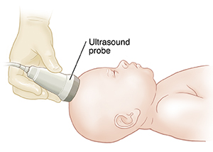
Healthcare providers can find PVL with an ultrasound (imaging test) of the head. But sometimes, signs of PVL can’t be seen with an ultrasound right away. So healthcare providers give babies at risk for PVL an ultrasound 4 to 8 weeks after birth. Magnetic resonance imaging (MRI) may also be used to find PVL.
 |
| An ultrasound of the head can show signs of PVL within a few weeks after birth. |
What are the long-term effects?
PVL may lead to problems with physical or mental development. How bad these problems are can vary. In some mild cases, healthcare providers find PVL on imaging tests. But the condition causes no symptoms. In these cases, the baby still needs to be checked from time to time for signs of problems. In the most severe cases, PVL can cause cerebral palsy or other serious physical and mental delays. Only time can tell how bad a child’s disability will be. If your child has PVL, they should be checked regularly by a developmental expert. This can help spot problems early so you and your child can get help early.
© 2000-2025 The StayWell Company, LLC. All rights reserved. This information is not intended as a substitute for professional medical care. Always follow your healthcare professional's instructions.